
#38: Carrot
Examining a carrot under a microscope unveils a mesmerizing world of intricate cellular structures. The microscope reveals a complex arrangement of cells that make up the carrot’s vibrant orange flesh. Individual cells appear elongated and tightly packed, forming the characteristic cylindrical shape of the vegetable. We can also see delicate vascular bundles, which transport water, nutrients, and sugars throughout the carrot.
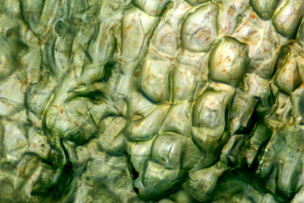
Additionally, microscopic examination shows the presence of tiny pigment-containing vesicles called chromoplasts, responsible for the carrot’s vibrant orange color. These chromoplasts contain pigments such as beta-carotene, which is a precursor to vitamin A. The microscopic view of a carrot offers a fascinating glimpse into the intricate composition of this nutritious root vegetable, highlighting the remarkable complexity that lies beneath its familiar appearance.